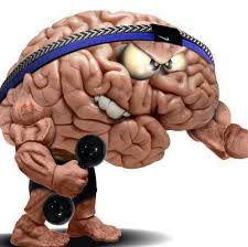
Brain helps to burn body fat

January 17, 2015

Brain burn

Jan 17: Not just doing bench presses and cardio, but your brain’s functioning is what helps your body lose fat and control weight. According to a recent research conducted at Monash University, our brain regulates and helps our body to burn fat. The researchers have explored how two hormones in the brain aid the body to burn fat.

How brain helps in losing weight?

Two hormones released by the brain namely, leptin and insulin jointly work towards converting white fat into brown fat. Leptin is an appetite suppressant produced in fat cells whereas insulin is generated in the pancreas in response to increased glucose levels in the blood. In fact the molecular mechanisms of these two hormones act in conjunction on a group of neurones to help the body burn fat through the nervous system.

According to the lead researcher Professor Tony Tiganis, from the Department of Biochemistry and Molecular Biology, the finding of the joint action of these two hormones might help in the burning of the excess fat in the body. Here are few exercises that could help you to boost your brain power.

So should you train your brain to burn more fat?

Going by what the researcher say, fat in the human body of the adults is usually deposited in adipocytes specialized cells that contain white fat. They also added that around the neck and shoulders, there is a second form of fat, which is made of brown adipocytes. These cells can be induced to burn the fat rather than storing it. The scientists revealed that leptin and insulin interact with proopiomelanocortin (POMC) neurons in the brain’s hypothalamus, making them send signals through the nervous system, stimulating the conversion of white fat into brown fat. This results in burning off of the excess fat in the human body.

How does the brain really work while helping to lose weight?

During the research the scientists managed to show that the process gets regulated in these neurons with the help of the enzymes known as phosphatases, which inhibit the actions of each of the hormones. When the levels of these inhibitors were decreased, the browning and burning of fat increased. According to the lead researcher, Tiganis, this important mechanism normally helps to maintain body weight but in the situation of diet-induced fatness this process goes haywire.

Comments

Add new comment

  • Coastaldigest.com reserves the right to delete or block any comments.
  • Coastaldigset.com is not responsible for its readers’ comments.
  • Comments that are abusive, incendiary or irrelevant are strictly prohibited.
  • Please use a genuine email ID and provide your name to avoid reject.
Agencies
April 4,2020

Washington D.C., Apr 4: While consuming a high-diet salt can result in high blood pressure, a recent study has revealed a link between salt-rich diet and weaker immune system.

The study was conducted under the leadership of the University Hospital Bonn, and the results were published in the journal Science Translational Medicine.

The research was conducted on mice that were fed a high-salt diet. Later, they were found to suffer from much more severe bacterial infections.

Human volunteers who consumed an additional six grams of salt per day also showed pronounced immune deficiencies.

The World Health Organization (WHO) has recommended a maximum amount of five grams of salt a day.

It corresponds approximately to one level teaspoon. In reality, however, many Germans exceed this limit considerably. 

Figures from the Robert Koch Institute suggest that on average men consume ten, and women more than eight grams a day.

This means that we reach for the salt shaker much more than is good for us. After all, sodium chloride, which is its chemical name, raises blood pressure and thereby increases the risk of heart attack or stroke.

"We have now been able to prove for the first time that excessive salt intake also significantly weakens an important arm of the immune system," said Prof. Dr. Christian Kurts from the Institute of Experimental Immunology at the University of Bonn.

This finding is unexpected, as some studies point in the opposite direction. For example, infections with certain skin parasites in laboratory animals heal significantly faster if these consume a high-salt diet.

The study also sheds light on the fact that the skin serves as a salt reservoir.

"Our results show that this generalization is not accurate," emphasized Katarzyna Jobin, lead author of the study.

The body keeps the salt concentration in the blood and in the various organs largely constant. Otherwise important biological processes would be impaired. The only major exception is the skin which functions as a salt reservoir of the body. This is why the additional intake of sodium chloride works so well for some skin diseases.

However, other parts of the body are not exposed to the additional salt consumed with food. Instead, it is filtered out by the kidneys and excreted in the urine.

"We examined volunteers who consumed six grams of salt in addition to their daily intake," said Prof. Kurts. This is roughly the amount contained in two fast-food meals, i.e. two burgers and two portions of French fries.

After one week, from the results, it showed that the immune cells coped much worse with bacteria after the test subjects had started to eat a high-salt diet.

In human volunteers, excessive salt intake also resulted in increased glucocorticoid levels.

Comments

Add new comment

  • Coastaldigest.com reserves the right to delete or block any comments.
  • Coastaldigset.com is not responsible for its readers’ comments.
  • Comments that are abusive, incendiary or irrelevant are strictly prohibited.
  • Please use a genuine email ID and provide your name to avoid reject.
Agencies
May 2,2020

Clinician-scientists have found that Irish patients admitted to hospital with severe coronavirus (COVID-19) infection are experiencing abnormal blood clotting that contributes to death in some patients.

The research team from the Royal College of Surgeons in Ireland found that abnormal blood clotting occurs in Irish patients with severe COVID-19 infection, causing micro-clots within the lungs.

According to the study, they also found that Irish patients with higher levels of blood clotting activity had a significantly worse prognosis and were more likely to require ICU admission.

"Our novel findings demonstrate that COVID-19 is associated with a unique type of blood clotting disorder that is primarily focussed within the lungs and which undoubtedly contributes to the high levels of mortality being seen in patients with COVID-19," said Professor James O'Donnell from St James's Hospital in Ireland.

In addition to pneumonia affecting the small air sacs within the lungs, the research team has also hundreds of small blood clots throughout the lungs.

This scenario is not seen with other types of lung infection and explains why blood oxygen levels fall dramatically in severe COVID-19 infection, the study, published in the British Journal of Haematology said.

"Understanding how these micro-clots are being formed within the lung is critical so that we can develop more effective treatments for our patients, particularly those in high-risk groups," O'Donnell said.

"Further studies will be required to investigate whether different blood-thinning treatments may have a role in selected high-risk patients in order to reduce the risk of clot formation," Professor O'Donnell added.

According to the study, emerging evidence also shows that the abnormal blood-clotting problem in COVID-19 results in a significantly increased risk of heart attacks and strokes.

As of Friday morning, the cases increased to 20,612 cases in Ireland, with 1,232 deaths so far, according to the Johns Hopkins University.

Comments

Add new comment

  • Coastaldigest.com reserves the right to delete or block any comments.
  • Coastaldigset.com is not responsible for its readers’ comments.
  • Comments that are abusive, incendiary or irrelevant are strictly prohibited.
  • Please use a genuine email ID and provide your name to avoid reject.
Agencies
July 24,2020

Colorado, Jul 24: A new study has found that physical stress in one's job may be associated with faster brain ageing and poorer memory.

Aga Burzynska, an assistant professor in the Department of Human Development and Family Studies, and her research team connected occupational survey responses with brain-imaging data from 99 cognitively normal older adults, age 60 to 79. They found that those who reported high levels of physical stress in their most recent job had smaller volumes in the hippocampus and performed poorer on memory tasks. The hippocampus is the part of the brain that is critical for memory and is affected in both normal ageing and in dementia.

Their findings were published this summer in Frontiers in Human Neuroscience under the research topic 'Work and Brain Health Across the Lifespan.'

"We know that stress can accelerate physical ageing and is the risk factor for many chronic illnesses," Burzynska said. "But this is the first evidence that occupational stress can accelerate brain and cognitive ageing."

She added that it is important to understand how occupational exposures affect the ageing of our brains.

"An average American worker spends more than eight hours at work per weekday, and most people remain in the workforce for over 40 years," Burzynska said. "By pure volume, occupational exposures outweigh the time we spend on leisure social, cognitive and physical activities, which protect our ageing minds and brains."

Physical demands at work

Burzynska explained that the association between "physical stress" and brain/memory were driven by physical demands at work. These included excessive reaching, or lifting boxes onto shelves, not necessarily aerobic activity. This is important because earlier work by Burzynska and her colleagues showed that leisure aerobic exercise is beneficial for brain health and cognition, from children to very old adults. Therefore, the researchers controlled for the effects of leisure physical activity and exercise.

As expected, leisure physical activity was associated with greater hippocampal volume, but the negative association with physical demands at work persisted.

"This finding suggests that physical demands at work may have parallel yet opposing associations with brain health," Burzynska explained. "Most interventions for postponing cognitive decline focus on leisure, not on your job. It's kind of unknown territory, but maybe future research can help us make some tweaks to our work environment for long-term cognitive health."

She added that the results could have important implications for society.

"Caring for people with cognitive impairment is so costly, on economic, emotional and societal levels," Burzynska said. "If we can support brain health earlier, in middle-aged workers, it could have an enormous impact."

The researchers considered and corrected for several other factors that could be related to work environment, memory and hippocampus, such as age, gender, brain size, educational level, job title, years in the occupation and general psychological stress.

One piece of the puzzle

"The research on this topic is so fragmented," Burzynska said. "One previous study linked mid-life managerial experience with greater hippocampus volume in older age. Another showed that taxi drivers had larger hippocampi than a city's bus drivers, presumably due to the need to navigate. In our study, job complexity and psychological stress at work were not related to hippocampal volume and cognition. Clearly, our study is just one piece of the puzzle, and further research is needed."

The magnetic resonance imaging (MRI) data used for the study was collected at the University of Illinois Urbana-Champaign between 2011 and 2014.

CSU researchers now can collect MRI data with the new 3T scanner at the University's Translational Medicine Institute.

With this new capability, Burzynska, along with Michael Thomas and Lorann Stallones of CSU's Department of Psychology, is launching a new project, "Impact of Occupational Exposures and Hazards on Brain and Cognitive Health Among Aging Agricultural Workers," which will involve collecting MRI brain scans and identifying risk and protective factors that could help the agricultural community age successfully. The project recently obtained funding as an Emerging Issues Short-Term Project from the High Plains Intermountain Center for Agricultural Health and Safety.

The Department of Human Development and Family Studies is part of CSU's College of Health and Human Sciences.

Comments

Add new comment

  • Coastaldigest.com reserves the right to delete or block any comments.
  • Coastaldigset.com is not responsible for its readers’ comments.
  • Comments that are abusive, incendiary or irrelevant are strictly prohibited.
  • Please use a genuine email ID and provide your name to avoid reject.