Study maps how brain recognise extensively varied faces at one go

December 27, 2016

Washington, Dec 27: Ever wondered how can you recognise whether your friend is happy or sad, at a glance? Also how can you recognise a friend, even if you haven't seen him/her in a decade?
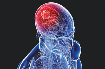
BrainAnswering to all these, a recent study finds out how the brain recognise familiar faces with efficiency and ease, despite extensive variation in how they appear.

Researchers at Carnegie Mellon University in the US are closer than ever before to understand the neural basis of facial identification.

The study, published in the journal of Proceedings of the National Academy of Sciences (PNAS), used highly sophisticated brain imaging tools and computational methods to measure the real-time brain processes that convert the appearance of a face into the recognition of an individual.

“Our results provide a step towards understanding the stages of information processing that begin when an image of a face first enters a person's eye and unfold over the next few hundred milliseconds, until the person is able to recognize the identity of the face," said study author Mark D. Vida.

To determine, how the brain rapidly distinguishes faces, they researchers scanned the brains of four people using magnetoencephalography (MEG).

MEG allowed them to measure ongoing brain activity throughout the brain on a millisecond-by-millisecond basis while the participants viewed images of 91 different individuals with two facial expressions each: happy and neutral.

The participants indicated that when they recognised that the same individual's face was repeated, regardless of expression.

The MEG scans allowed the researchers to map out, for each of many points in time, which parts of the brain encode appearance-based information and which encode identity-based information.

The team also compared the neural data to behavioral judgments of the face images from humans, whose judgments were based mainly on identity-based information.

Then, they validated the results by comparing the neural data to the information present in different parts of a computational simulation of an artificial neural network that was trained to recognise individuals from the same face images.

“Combining the detailed timing information from MEG imaging with computational models of how the visual system works has the potential to provide insight into the real-time brain processes underlying many other abilities beyond face recognition," said another researcher David C. Plaut.

The researchers are hopeful that the findings might be used in the near future to locate the exact point at which the visual perception system breaks down in different disorders and injuries, ranging from developmental dyslexia to prosopagnosia or face blindness.

Comments

Add new comment

  • Coastaldigest.com reserves the right to delete or block any comments.
  • Coastaldigset.com is not responsible for its readers’ comments.
  • Comments that are abusive, incendiary or irrelevant are strictly prohibited.
  • Please use a genuine email ID and provide your name to avoid reject.
Agencies
May 2,2020

Clinician-scientists have found that Irish patients admitted to hospital with severe coronavirus (COVID-19) infection are experiencing abnormal blood clotting that contributes to death in some patients.

The research team from the Royal College of Surgeons in Ireland found that abnormal blood clotting occurs in Irish patients with severe COVID-19 infection, causing micro-clots within the lungs.

According to the study, they also found that Irish patients with higher levels of blood clotting activity had a significantly worse prognosis and were more likely to require ICU admission.

"Our novel findings demonstrate that COVID-19 is associated with a unique type of blood clotting disorder that is primarily focussed within the lungs and which undoubtedly contributes to the high levels of mortality being seen in patients with COVID-19," said Professor James O'Donnell from St James's Hospital in Ireland.

In addition to pneumonia affecting the small air sacs within the lungs, the research team has also hundreds of small blood clots throughout the lungs.

This scenario is not seen with other types of lung infection and explains why blood oxygen levels fall dramatically in severe COVID-19 infection, the study, published in the British Journal of Haematology said.

"Understanding how these micro-clots are being formed within the lung is critical so that we can develop more effective treatments for our patients, particularly those in high-risk groups," O'Donnell said.

"Further studies will be required to investigate whether different blood-thinning treatments may have a role in selected high-risk patients in order to reduce the risk of clot formation," Professor O'Donnell added.

According to the study, emerging evidence also shows that the abnormal blood-clotting problem in COVID-19 results in a significantly increased risk of heart attacks and strokes.

As of Friday morning, the cases increased to 20,612 cases in Ireland, with 1,232 deaths so far, according to the Johns Hopkins University.

Comments

Add new comment

  • Coastaldigest.com reserves the right to delete or block any comments.
  • Coastaldigset.com is not responsible for its readers’ comments.
  • Comments that are abusive, incendiary or irrelevant are strictly prohibited.
  • Please use a genuine email ID and provide your name to avoid reject.
Agencies
June 19,2020

While coughing, fever and difficulty in breathing are common symptoms of COVID-19, a new case study has found that pink eye is also a reason to be tested for the disease.

The study, published in the Canadian Journal of Ophthalmology, determined that conjunctivitis and keratoconjunctivitis can also be primary symptoms of COVID-19.

The researchers noted that in March, a 29-year-old woman arrived at the Royal Alexandra Hospital's Eye Institute of Alberta with a severe case of conjunctivitis and minimal respiratory symptoms.

After the patient had undergone several days of treatment with little improvement -- and after it had been determined that the woman had recently returned home from Asia -- a resident ordered a COVID-19 test.

The test came back positive, according to the researchers.

"What is interesting in this case, and perhaps very different to how it had been recognised at that specific time, was that the main presentation of the illness was not a respiratory symptom. It was the eye," said Carlos Solarte, an assistant professor at the University of Alberta in Canada.

"There was no fever and no cough, so we weren't led to suspect COVID-19 at the beginning. We didn't know it could present primarily with the eye and not with the lungs," Solarte said.

Academic studies at the outset of the pandemic identified conjunctivitis as a secondary symptoms in about 10 to 15 per cent of COVID-19 cases, he said.

Since then, scientists have gained greater knowledge of how the virus can transmit through and affect the body's mucous membrane system, of which the conjunctiva -- the clear, thin membrane that covers the front surface of the eye -- is an extension.

While the finding provides important new health information for the public, it also makes eye exams more complicated for ophthalmologists and staff, the researchers noted.

"The patient in this case eventually recovered well without any issues. But several of the residents and staff who were in close contact with the patient had to be under quarantine," said Solarte.

"Fortunately, none who were involved in her care also tested positive," he said.

Patients coming into an eye clinic with conjunctivitis and keratoconjunctivitis are now treated as potential cases of COVID-19 and extra precautions are taken by staff, according to the researchers.

Comments

Add new comment

  • Coastaldigest.com reserves the right to delete or block any comments.
  • Coastaldigset.com is not responsible for its readers’ comments.
  • Comments that are abusive, incendiary or irrelevant are strictly prohibited.
  • Please use a genuine email ID and provide your name to avoid reject.
Agencies
July 3,2020

Apart from the many benefits of doing exercise, new research has now found that exercise can slow down or prevent the development of macular degeneration and may benefit other common causes of vision loss, such as glaucoma and diabetic retinopathy.

The new study from the University of Virginia School of Medicine found that exercise reduced the harmful overgrowth of blood vessels in the eyes of lab mice by up to 45 per cent. This tangle of blood vessels is a key contributor to macular degeneration and several other eye diseases.

The study represents the first experimental evidence showing that exercise can reduce the severity of macular degeneration, a leading cause of vision loss, the scientists report. Ten million Americans are estimated to have the condition.

"There has long been a question about whether maintaining a healthy lifestyle can delay or prevent the development of macular degeneration. The way that question has historically been answered has been by taking surveys of people, asking them what they are eating and how much exercise they are performing," said researcher Bradley Gelfand, PhD, of UVA's Center for Advanced Vision Science.

"That is basically the most sophisticated study that has been done. The problem with that is that people are notoriously bad self-reporters ... and that can lead to conclusions that may or not be true. This [study] offers hard evidence from the lab for the very first time," Gelfand added.

Enticingly, the research found that the bar for receiving the benefits from exercise was relatively low - more exercise didn't mean more benefit.

"Mice are kind of like people in that they will do a spectrum of exercise. As long as they had a wheel and ran on it, there was a benefit. The benefit that they obtained is saturated at low levels of exercise," Gelfand said.

An initial test comparing mice that voluntarily exercised versus those that did not found that exercise reduced the blood vessel overgrowth by 45%. A second test, to confirm the findings, found a reduction of 32 per cent.

The scientists aren't certain exactly how exercise is preventing the blood vessel overgrowth. There could be a variety of factors at play, they say, including increased blood flow to the eyes.

Gelfand, of UVA's Department of Ophthalmology and Department of Biomedical Engineering, noted that the onset of vision loss is often associated with a decrease in exercise.

"It is fairly well known that as people's eyes and vision deteriorate, their tendency to engage in physical activity also goes down. It can be a challenging thing to study with older people. ... How much of that is one causing the other?" he said.
The researchers already have submitted grant proposals in hopes of obtaining funding to pursue their findings further.

"The next step is to look at how and why this happens, and to see if we can develop a pill or method that will give you the benefits of exercise without having to exercise," Gelfand said.

He explained, "We're talking about a fairly elderly population [of people with macular degeneration], many of whom may not be capable of conducting the type of exercise regimen that may be required to see some kind of benefit." (He urged people to consult their doctors before beginning any aggressive exercise program.)
Gelfand, a self-described couch potato, disclosed a secret motivation for the research: "One reason I wanted to do this study was sort of selfish. I was hoping to find some reason not to exercise," he joked. "It turned out exercise really is good for you."

Comments

Add new comment

  • Coastaldigest.com reserves the right to delete or block any comments.
  • Coastaldigset.com is not responsible for its readers’ comments.
  • Comments that are abusive, incendiary or irrelevant are strictly prohibited.
  • Please use a genuine email ID and provide your name to avoid reject.