Study maps how brain recognise extensively varied faces at one go

December 27, 2016

Washington, Dec 27: Ever wondered how can you recognise whether your friend is happy or sad, at a glance? Also how can you recognise a friend, even if you haven't seen him/her in a decade?

BrainAnswering to all these, a recent study finds out how the brain recognise familiar faces with efficiency and ease, despite extensive variation in how they appear.

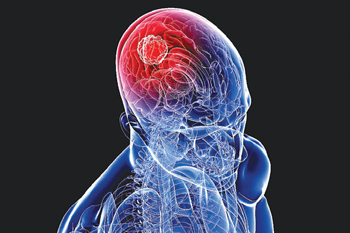
Researchers at Carnegie Mellon University in the US are closer than ever before to understand the neural basis of facial identification.

The study, published in the journal of Proceedings of the National Academy of Sciences (PNAS), used highly sophisticated brain imaging tools and computational methods to measure the real-time brain processes that convert the appearance of a face into the recognition of an individual.

“Our results provide a step towards understanding the stages of information processing that begin when an image of a face first enters a person's eye and unfold over the next few hundred milliseconds, until the person is able to recognize the identity of the face," said study author Mark D. Vida.

To determine, how the brain rapidly distinguishes faces, they researchers scanned the brains of four people using magnetoencephalography (MEG).

MEG allowed them to measure ongoing brain activity throughout the brain on a millisecond-by-millisecond basis while the participants viewed images of 91 different individuals with two facial expressions each: happy and neutral.

The participants indicated that when they recognised that the same individual's face was repeated, regardless of expression.

The MEG scans allowed the researchers to map out, for each of many points in time, which parts of the brain encode appearance-based information and which encode identity-based information.

The team also compared the neural data to behavioral judgments of the face images from humans, whose judgments were based mainly on identity-based information.

Then, they validated the results by comparing the neural data to the information present in different parts of a computational simulation of an artificial neural network that was trained to recognise individuals from the same face images.

“Combining the detailed timing information from MEG imaging with computational models of how the visual system works has the potential to provide insight into the real-time brain processes underlying many other abilities beyond face recognition," said another researcher David C. Plaut.

The researchers are hopeful that the findings might be used in the near future to locate the exact point at which the visual perception system breaks down in different disorders and injuries, ranging from developmental dyslexia to prosopagnosia or face blindness.

Comments

Add new comment

  • Coastaldigest.com reserves the right to delete or block any comments.
  • Coastaldigset.com is not responsible for its readers’ comments.
  • Comments that are abusive, incendiary or irrelevant are strictly prohibited.
  • Please use a genuine email ID and provide your name to avoid reject.
News Network
February 22,2020

Feb 22: The subjective feeling of well-being experienced by many people with the practice of meditation is associated with specific changes in the brain, according to a study which may lead to better clinical recommendations of the practice.

The study, published in the journal Brain and Cognition, examined the effects of the technique known as Transcendental Meditation (TM), which consists of the silent repetition of a meaningless sound.

In the study, the researchers from the IMT School for Advanced Studies Lucca in Italy, enrolled 34 healthy young volunteers and divided them in two groups.

They said the first group practised TM 40 minutes per day in two sessions of 20 minutes each, one in the morning and the other in the evening.

The second group, the scientists said, did not change its daily routine.

Using questionnaires, they also measured the anxiety and stress levels of all the participants at the beginning of the study, as well as the subjects' ability to manage stressful situations.

According to the researchers, the participants were also subjected to a functional magnetic resonance imaging (fMRI) brain scan, in order to measure the organ's activity at rest, and changes in the excitation among different cerebral areas.

They repeated the tests after three months, at the end of the study.

According to the study, the levels of anxiety and stress perceived by the subjects who followed the meditation program were significantly reduced in comparison with those of the volunteers who did not practice TM.

"Magnetic resonance imaging also shows that the reduction of anxiety levels is associated with specific changes in the connectivity between different cerebral areas, such as precuneus, left parietal lobe and insula, which all have an important role in the modulation of emotions and inner states," said study co-author Giulia Avvenuti from the IMT School for Advanced Studies Lucca.

"In the control group, instead, none of these changes was observed. The fact that Transcendental Meditation has measurable effects on the 'dialogue' between brain structures involved in the modulation of affective states opens new perspectives for the understanding of brain-mind relationships," said Pietro Pietrini, IMT School's Director, and co-author of the study.

"It also extends the results of recent research suggesting that drugs therapies and psychotherapy leverage on the same biological mechanism," Pietrini said.

According to the researchers, even a few months of practice of TM can have positive effects which can be correlated with measurable changes in the brain.

Comments

Add new comment

  • Coastaldigest.com reserves the right to delete or block any comments.
  • Coastaldigset.com is not responsible for its readers’ comments.
  • Comments that are abusive, incendiary or irrelevant are strictly prohibited.
  • Please use a genuine email ID and provide your name to avoid reject.
Agencies
June 29,2020

Washington DC, Jun 29: Young children with narrow retinal artery diameters were more likely to develop higher blood pressure, and children with higher blood pressure levels were more likely to develop retinal microvascular impairment during early childhood, according to a new study.

The first study to show this connection in children was published today in Hypertension, an American Heart Association journal.

High blood pressure, the main risk factor for the development of cardiovascular disease (CVD), can manifest as early as childhood, and the prevalence of high blood pressure among children continues to rise. In previous studies, analysis of blood vessels in the retina has shown promise as a predictor of CVD risk among adults. In the study titled, "Retinal Vessel Diameters and Blood Pressure Progression in Children," researchers sought to predict the development of high blood pressure in children over four years based on retinal blood vessel measurements.

"Hypertension continues as the main risk factor for the development of cardiovascular diseases and mortality," says Henner Hanssen, M.D., the study's lead author and a professor in the department of sport, exercise and health at the University of Basel in Switzerland. 

"Primary prevention strategies are needed to focus on screening retinal microvascular health and blood pressure in young children in order to identify those at increased risk of developing hypertension. The earlier we can provide treatment and implement lifestyle changes to reduce hypertension, the greater the benefit for these children."

Researchers screened 262 children ages six to eight from 26 schools in Basel, Switzerland, in 2014, for baseline blood pressure and retinal arterial measurements. Both measures were taken again in 2018. Blood pressure measurements at both baseline and follow-up were performed in a sitting position after a minimum of five minutes of rest and were categorized based on the American Academy of Pediatrics' blood pressure guidelines. These guidelines utilize the same measurements as the American Heart Association/American College of Cardiology 2017 Guideline for the Prevention, Detection, Evaluation, and Management of High Blood Pressure in Adults.

Results from the analysis indicate: children with narrower retinal vessel diameters at baseline developed higher systolic blood pressure at follow-up; retinal vessel diameters could explain 29 -31 per cent of the changes in systolic blood pressure progression between 2014 and 2018; children with higher blood pressure levels at baseline developed significantly narrower arteriolar diameters at follow-up, depending on weight and cardiorespiratory fitness; and initial blood pressure measures explained 66-69 per cent of the change in retinal arteriolar diameter from baseline to follow-up.

"Early childhood assessments of retinal microvascular health and blood pressure monitoring can improve cardiovascular risk classification. Timely primary prevention strategies for children at risk of developing hypertension could potentially counteract its growing burden among both children and adults," said Hanssen.

Researchers noted limitations of their study include that they could not confirm blood pressure measurements over a single 24-hour period, so they would not account for "white coat" hypertension, a condition where patients have high blood pressure readings when measured in a medical setting.

Developmental stage including puberty status of each child was not accounted for in the study, as well as genetic factors or birth weight - variables that could impact blood pressure development and microvascular health.

In addition, reference values for appropriate retinal vessel diameters in children do not currently exist, so future studies are needed to determine age-related normal values during childhood.

Comments

Add new comment

  • Coastaldigest.com reserves the right to delete or block any comments.
  • Coastaldigset.com is not responsible for its readers’ comments.
  • Comments that are abusive, incendiary or irrelevant are strictly prohibited.
  • Please use a genuine email ID and provide your name to avoid reject.
Agencies
January 26,2020

High-protein diets may help people lose weight and build muscle, but there is a downside to it --a greater heart attack risk. Researchers now report that high-protein diets boost artery-clogging plaque.

The research in mice showed that high-protein diets spur unstable plaque -- the kind most prone to rupturing and causing blocked arteries.

More plaque buildup in the arteries, particularly if it's unstable, increases the risk of heart attack.

"There are clear weight-loss benefits to high-protein diets, which has boosted their popularity in recent years," said senior author Babak Razani, associate professor at Washington University School of Medicine in St. Louis, Missouri.

"But animal studies and some large epidemiological studies in people have linked high dietary protein to cardiovascular problems. We decided to take a look at whether there is truly a causal link between high dietary protein and poorer cardiovascular health," Razani added.

The researchers studied mice who were fed a high-fat diet to deliberately induce atherosclerosis, or plaque buildup in the arteries.

Some of the mice received a high-fat diet that was also high in protein. And others were fed a high-fat, low-protein diet for comparison.

The mice on the high-fat, high-protein diet developed worse atherosclerosis -- about 30 per cent more plaque in the arteries -- than mice on the high-fat, normal-protein diet, despite the fact that the mice eating more protein did not gain weight, unlike the mice on the high-fat, normal-protein diet.

"A couple of a scoop of protein powder in a milkshake or smoothie adds something like 40 grams of protein -- almost equivalent to the daily recommended intake," Razani said.

"To see if protein has an effect on cardiovascular health, we tripled the amount of protein that the mice receive in the high-fat, high-protein diet -- keeping the fat constant. Protein went from 15 per cent to 46 per cent of calories for these mice".

Plaque contains a mix of fat, cholesterol, calcium deposits and dead cells. Past work by Razani's team and other groups has shown that immune cells called macrophages work to clean up plaque in the arteries.

But the environment inside plaque can overwhelm these cells, and when such cells die, they make the problem worse, contributing to plaque buildup and increasing plaque complexity.

"In mice on the high-protein diet, their plaques were a macrophage graveyard," Razani informed.

To understand how high dietary protein might increase plaque complexity, Razani and his colleagues also studied the path protein takes after it has been digested -- broken down into its original building blocks, called amino acids.

"This study is not the first to show a telltale increase in plaque with high-protein diets, but it offers a deeper understanding of the impact of high protein with the detailed analysis of the plaques," said Razani.

"This work not only defines the critical processes underlying the cardiovascular risks of dietary protein but also lays the groundwork for targeting these pathways in treating heart disease," he added.

Comments

Add new comment

  • Coastaldigest.com reserves the right to delete or block any comments.
  • Coastaldigset.com is not responsible for its readers’ comments.
  • Comments that are abusive, incendiary or irrelevant are strictly prohibited.
  • Please use a genuine email ID and provide your name to avoid reject.