'Muscles, not brain, may be behind sleep disorders'

Agencies
August 6, 2017

Houston, Aug 6: The answer to combat sleep disorders may not be in the brain, say scientists who have found that a protein present in the muscles can lessen the effects of sleep loss.

Researchers from University of Texas Southwestern in the US demonstrated how a circadian clock protein in the muscle - BMAL1 - regulates the length and manner of sleep in mice.

The surprising revelation challenges the widely accepted notion that the brain controls all aspects of sleep, they said.

The team found that while the protein's presence or absence in the brain had little effect on sleep recovery, mice with higher levels of BMAL1 in their muscles recovered from sleep deprivation more quickly.

Removing BMAL1 from the muscle severely disrupted normal sleep, leading to an increased need for sleep, deeper sleep, and a reduced ability to recover, researchers said.

"These studies show that factors in muscles can signal to the brain to influence sleep. If similar pathways exist in people, this would provide new drug targets for the treatment of sleep disorders,” said Joseph S Takahashi, Chairman of Neuroscience at UT Southwestern Medical Center.

"This finding is completely unexpected and changes the ways we think sleep is controlled," Takahashi added. The study was published in the journal eLife.

Comments

Add new comment

  • Coastaldigest.com reserves the right to delete or block any comments.
  • Coastaldigset.com is not responsible for its readers’ comments.
  • Comments that are abusive, incendiary or irrelevant are strictly prohibited.
  • Please use a genuine email ID and provide your name to avoid reject.
News Network
February 26,2020

New York, Feb 26:  A new wearable sensor that works in conjunction with artificial intelligence (AI) technology could help doctors remotely detect critical changes in heart failure patients days before a health crisis occurs, says a study.

The researchers said the system could eventually help avert up to one in three heart failure readmissions in the weeks following initial discharge from the hospital and help patients sustain a better quality of life.

"This study shows that we can accurately predict the likelihood of hospitalisation for heart failure deterioration well before doctors and patients know that something is wrong," says the study's lead author Josef Stehlik from University of Utah in the US.

"Being able to readily detect changes in the heart sufficiently early will allow physicians to initiate prompt interventions that could prevent rehospitalisation and stave off worsening heart failure," Stehlik added.

According to the researchers, even if patients survive, they have poor functional capacity, poor exercise tolerance and low quality of life after hospitalisations.

"This patch, this new diagnostic tool, could potentially help us prevent hospitalizations and decline in patient status," Stehlik said.

For the findings, published in the journal Circulation: Heart Failure, the researchers followed 100 heart failure patients, average age 68, who were diagnosed and treated at four veterans administration (VA) hospitals in Utah, Texas, California, and Florida.

After discharge, participants wore an adhesive sensor patch on their chests 24 hours a day for up to three months.

The sensor monitored continuous electrocardiogram (ECG) and motion of each subject.

This information was transmitted from the sensor via Bluetooth to a smartphone and then passed on to an analytics platform, developed by PhysIQ, on a secure server, which derived heart rate, heart rhythm, respiratory rate, walking, sleep, body posture and other normal activities.

Using artificial intelligence, the analytics established a normal baseline for each patient. When the data deviated from normal, the platform generated an indication that the patient's heart failure was getting worse.

Overall, the system accurately predicted the impending need for hospitalization more than 80 per cent of the time.

On average, this prediction occurred 10.4 days before a readmission took place (median 6.5 days), the study said.

Comments

Add new comment

  • Coastaldigest.com reserves the right to delete or block any comments.
  • Coastaldigset.com is not responsible for its readers’ comments.
  • Comments that are abusive, incendiary or irrelevant are strictly prohibited.
  • Please use a genuine email ID and provide your name to avoid reject.
Dr G K Sudhakar Reddy
August 4,2020

Being overweight or obese is now recognised as a serious cause of ill health and disability. There is a significant positive association between orthopaedic disorders and the level of obesity causing pain, deformity and difficulty in walking.

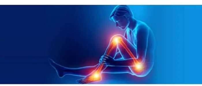
Excess body weight accumulation increases pressure on joints, particularly the hips, knees and ankles.

Here are a few type of  arthritis:

Osteoarthritis

It is a condition of damage/ wear and tear of the joint lining or cartilage. Obesity triggers this by loading excessive weight on the weight bearing joints like the knee and the hip. 

Knee Osteoarthritis

This is the most common arthritis especially in the Indian subcontinent.

While walking, an individual exerts 3 to 6 times pressure that of the body weight on the weight-bearing knee joint, which means in an obese with excess body weight, larger forces are exerted, which lead to higher risk of deterioration of cartilage.

In addition, there are excessive fat tissues that produce hormones and other factors that affect the joint cartilage metabolism and cause inflammation of the joints giving rise to joint pathology.  Leptin is one of the hormones causing knee osteoarthritis. 

Hip osteoarthritis

The force exerted across the hip is 3 times that of body weight. Hip osteoarthritis is caused by factors such as joint injury, increasing age and being overweight.    

Hand osteoarthritis

The observation that obese individual has a higher risk in having hand osteoarthritis has led to a hypothesis that the metabolic effect produced by fat tissue is the underlying factor. 

Osteoporosis

It is a progressive bone condition of decrease in bone mass and density (Bone Mineral Density or BMD) which can lead to an increased risk of fracture. Recent research suggests that obesity may accelerate bone loss. It is the amount of muscle mass which is seen in an active person, which accounts for bone strengthening effects and not due to the fat seen in a heavy person.

Low back pain

Low back pain from degenerative disc disease of the lumbar spine is one of the most disabling conditions in the community and overweight and obesity have the strongest association with seeking care for low back pain.

Managing Hip and Knee Osteoarthritis

Life style changes

If one is overweight, try to lose weight by doing more physical activity and eating a healthier diet. Regular exercise keeps you active and mobile and builds up muscle, thereby strengthening the joints and can improve symptoms. 

Pain Killers

Painkillers help with pain and stiffness for short term. They don’t affect the arthritis itself and won’t repair the damage to your joint. Creams and gels can be applied directly onto painful joints.

Nutritional Supplements

Glucosamine and chondroitin are nutritional supplements. Animal studies have found that glucosamine can both delay the breakdown of and repair damaged cartilage. However, there is insufficient evidence to support the use of glucosamine in humans and one can expect only a mild-to-moderate reduction in pain

Joint injections

If pain from osteoarthritis is severe joint steroid injections are injected into the joints that can reduces swelling and pain. The injections can start working within a day or so and may improve pain for several weeks or months. 

Hyaluronic acid injections, which help to lubricate your knee joint also give short term relief. In early stages. Stem cell treatment or cartilage regeneration procedures are being tried in young people with small defects, however it is still experimental and lacks long term evidence.

Surgery

May be recommended if you have severe pain or mobility problems.

Arthroscopy

If one has frequent painful locking/stiffening episodes especially in the knee joint, an operation to wash out loose fragments of bone and other tissue as joint can be performed by a minimally invasive key hole procedure called Arthroscopy.

Arthrodesis

If hip or knee replacement is not suitable, especially in young people who do heavy manual work, one can consider an operation known as an arthrodesis, which fuses your joint in a permanent position. This means that your joint will be stronger and much less painful, although you will no longer be able to move it.

Osteotomy

In young, active people in whom a knee joint replacement would fail due to excessive use one can consider an operation called an osteotomy. This involves adding or removing a small section of bone either above or below your knee joint.  This helps realign your knee so your weight is no longer focused on the damaged part of your knee. An osteotomy can relieve your symptoms of osteoarthritis, although you may still need knee replacement surgery eventually as you grow old

Joint replacement surgery

Joint replacement therapy is most commonly carried out to replace hip and knee joints. It involves replacing a damaged, worn or diseased joint with an artificial joint made of special plastics and metal.

For most people, a replacement hip or knee will last for at least 20 years, especially if it is cared for properly and not put under too much strain.

Dr G K Sudhakar Reddy is a Sr Consultant Orthopaedic Surgeon at Citizens Speciality Hospitals, Hyderabad

Comments

Add new comment

  • Coastaldigest.com reserves the right to delete or block any comments.
  • Coastaldigset.com is not responsible for its readers’ comments.
  • Comments that are abusive, incendiary or irrelevant are strictly prohibited.
  • Please use a genuine email ID and provide your name to avoid reject.
Agencies
June 23,2020

The record levels of new daily COVID-19 cases are due to the fact that the pandemic is peaking in a number of big countries at the same time and reflect a change in the virus' global activity, the World Health Organisation said.

At a media briefing on Monday, WHO's emergencies chief Dr Michael Ryan said that the numbers are increasing because the epidemic is developing in a number of populous countries at the same time.

Some countries have attributed their increased caseload to more testing, including India and the US But Ryan dismissed that explanation.

We do not believe this is a testing phenomenon, he said, noting that numerous countries have also noted marked increases in hospital admissions and deaths neither of which cannot be explained by increased testing.

There definitely is a shift in that the virus is now very well established, Ryan said. The epidemic is now peaking or moving towards a peak in a number of large countries.

He added the situation was definitely accelerating in a number of countries, including the US and others in South Asia, the Middle East and Africa.

Comments

Add new comment

  • Coastaldigest.com reserves the right to delete or block any comments.
  • Coastaldigset.com is not responsible for its readers’ comments.
  • Comments that are abusive, incendiary or irrelevant are strictly prohibited.
  • Please use a genuine email ID and provide your name to avoid reject.