Soon, a blood test to detect shaken baby syndrome

April 11, 2017

Washington, Apr 11: A team of scientists has come up with a new medical test that may help identify infants who may have had bleeding of the brain as a result of abusive head trauma, sometimes referred to as shaken baby syndrome.

bloodDeveloped by the researchers at Children's Hospital of Pittsburgh of UPMC and University of Pittsburgh School of Medicine, the serum-based test, which needs to be validated in a larger population and receive regulatory approval before being used in clinical practice, would be the first of its kind to be used to detect acute intracranial hemorrhage or bleeding of the brain.

Infants who test positive would then have further evaluation via brain imaging to determine the source of the bleeding. "Abusive head trauma (AHT) is the leading cause of death from traumatic brain injury in infants and the leading cause of death from physical abuse in the United States," said senior author Rachel Berger.

However, approximately 30 percent of AHT diagnoses are missed when caretakers provide inaccurate histories or when infants have nonspecific symptoms such as vomiting or fussiness. Missed diagnoses can be catastrophic as AHT can lead to permanent brain damage and even death.

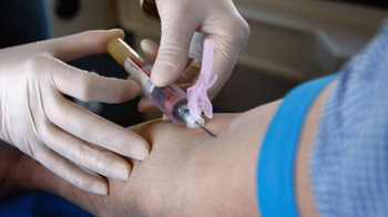
The researchers collaborated with Axela, a Canadian molecular diagnostics company, to develop a sensitive test that could reduce the chances of a missed diagnosis by using a combination of three biomarkers along with a measure of the patient's level of hemoglobin, the protein that carries oxygen in blood.

Axela's automated testing system allowed the researchers to measure multiple biomarkers simultaneously using an extremely small amount of blood, an important characteristic of a test designed to be used in infants.

The test correctly detected acute intracranial hemorrhage because of abusive head trauma approximately 90 percent of the time, a much higher rate than the sensitivity of clinical judgement, which is approximately 70 percent.

"The test is not intended to replace clinical judgement, which is crucial," said Berger. "Rather, we believe that it can supplement clinical evaluation and in cases where symptoms may be unclear, help physicians make a decision about whether an infant needs brain imaging."

The specificity of the test or the ability to correctly identify an infant without bleeding of the brain who would not require further evaluation was 48 percent. The researchers aimed for the test to be highly sensitive rather than maximizing accuracy, since missing a diagnosis has more serious consequences than performing brain imaging in babies without the condition.

"This study illustrates the benefits of being able to perform highly sensitive tests at the point of care," said co-author Paul Smith. The science behind the test is described in JAMA Pediatrics.

Comments

Add new comment

  • Coastaldigest.com reserves the right to delete or block any comments.
  • Coastaldigset.com is not responsible for its readers’ comments.
  • Comments that are abusive, incendiary or irrelevant are strictly prohibited.
  • Please use a genuine email ID and provide your name to avoid reject.
News Network
February 22,2020

Feb 22: The subjective feeling of well-being experienced by many people with the practice of meditation is associated with specific changes in the brain, according to a study which may lead to better clinical recommendations of the practice.

The study, published in the journal Brain and Cognition, examined the effects of the technique known as Transcendental Meditation (TM), which consists of the silent repetition of a meaningless sound.

In the study, the researchers from the IMT School for Advanced Studies Lucca in Italy, enrolled 34 healthy young volunteers and divided them in two groups.

They said the first group practised TM 40 minutes per day in two sessions of 20 minutes each, one in the morning and the other in the evening.

The second group, the scientists said, did not change its daily routine.

Using questionnaires, they also measured the anxiety and stress levels of all the participants at the beginning of the study, as well as the subjects' ability to manage stressful situations.

According to the researchers, the participants were also subjected to a functional magnetic resonance imaging (fMRI) brain scan, in order to measure the organ's activity at rest, and changes in the excitation among different cerebral areas.

They repeated the tests after three months, at the end of the study.

According to the study, the levels of anxiety and stress perceived by the subjects who followed the meditation program were significantly reduced in comparison with those of the volunteers who did not practice TM.

"Magnetic resonance imaging also shows that the reduction of anxiety levels is associated with specific changes in the connectivity between different cerebral areas, such as precuneus, left parietal lobe and insula, which all have an important role in the modulation of emotions and inner states," said study co-author Giulia Avvenuti from the IMT School for Advanced Studies Lucca.

"In the control group, instead, none of these changes was observed. The fact that Transcendental Meditation has measurable effects on the 'dialogue' between brain structures involved in the modulation of affective states opens new perspectives for the understanding of brain-mind relationships," said Pietro Pietrini, IMT School's Director, and co-author of the study.

"It also extends the results of recent research suggesting that drugs therapies and psychotherapy leverage on the same biological mechanism," Pietrini said.

According to the researchers, even a few months of practice of TM can have positive effects which can be correlated with measurable changes in the brain.

Comments

Add new comment

  • Coastaldigest.com reserves the right to delete or block any comments.
  • Coastaldigset.com is not responsible for its readers’ comments.
  • Comments that are abusive, incendiary or irrelevant are strictly prohibited.
  • Please use a genuine email ID and provide your name to avoid reject.
Agencies
January 16,2020

Thiruvananthapuram, Jan 16: Kerala Tourism on Wednesday shared a recipe of a popular meat dish in the Central Travancore region of Kerala, Beef Ularthiyathu, which is a special delicacy in the region.

Taking to its Twitter handle, the Kerala Tourism wrote, "Tender chunks of beef, slow-roasted with aromatic spices, coconut pieces, and curry leaves. A recipe for the most classic dish, Beef Ularthiyathu, the stuff of legends, from the land of spices, Kerala."

The State Tourism also shared the recipe of the delicacy with Twitteratis.

The tweet which has garnered 3.5 thousand likes so far had received a mixed response

While some said "beef is not Kerala's culture", others termed the recipe 'a match made in heaven".

Dr Vireandta Jilowa wrote, "Surprised to see it, that beef is being consumed despite BJP government in the Centre."

"We are not slaves of BJP at the Centre....people eat whatever they like in this state, including beef, pork, mutton and fish," another user Tatheesh Vijayakumar wrote.

In 2017, The Minister for Environment, Forest and Climate Change Harsh Vardhan had ordered that the ministry has notified the Prevention of Cruelty to Animals (Regulation of Livestock Markets) Rules, 2017 to ensure that the sale of cattle is not meant for slaughter purposes.

Regulating animal trade is a state business, but animal welfare is a central subject.

In lieu of this, there was widespread opposition of the order, with many states openly denying accepting the notification.

Porotta and Kappa biriyani with beef are counted as delicacies by Keralites. 

Also Read: The Art of Prepping Meat

Comments

Add new comment

  • Coastaldigest.com reserves the right to delete or block any comments.
  • Coastaldigset.com is not responsible for its readers’ comments.
  • Comments that are abusive, incendiary or irrelevant are strictly prohibited.
  • Please use a genuine email ID and provide your name to avoid reject.
Agencies
January 25,2020

Washington D.C., Jan 25: A new study conducted by a team of researchers reveals why individuals who have a history of early life adversity (ELA) are disproportionately prone to opioid addiction.

The study conducted examined how early adversities interact with factors such as increased access to opioids to directly influence brain development and function, causing a higher potential for opioid addiction.

The study was lead by UCI researchers and was published in Molecular Psychiatry.

Tallie Z. Baram, MD, PhD, the Danette Shepard Chair in Neurological Sciences at the UCI School of Medicine and one of the senior researchers for the study, was on the take that the widely known factor genetics that plays major role in addiction vulnerability, cannot be solely held responsible for the recent rise in opioid abuse.

To further clarify, the researchers simulated ELA in rats by limiting bedding and nesting materials during a short, postnatal period of time.

In female rats, this led to striking opioid addiction-like characteristics including an increased relapse- behaviour, for example.

As observed in addicted humans, the rats were willing to work very hard (pay a very high price) to obtain the drug.

Baram said: "Ultimately, we found that conditions during sensitive developmental periods can lead to vulnerability to the addictive effects of opioid drugs, especially in females, which is consistent with the prevalence of ELA in heroin-addicted women."

These findings can be used to highlight the importance given to sex differences in future ELA-related studies on opioid addiction, and in future prevention or intervention strategies being developed to address the growing opioid crisis.

The study conducted examined how early adversities interact with factors such as increased access to opioids to directly influence brain development and function, causing a higher potential for opioid addiction.

The study was lead by UCI researchers and was published in Molecular Psychiatry.

The study found that unpredictable, fragmented early life environments may lead to abnormal maturation of certain brain circuits, which profoundly impacts brain function and persists into adolescence and adulthood.

Tallie Z. Baram, MD, PhD, the Danette Shepard Chair in Neurological Sciences at the UCI School of Medicine and one of the senior researchers for the study, was on the take that the widely known factor genetics that plays major role in addiction vulnerability, cannot be solely held responsible for the recent rise in opioid abuse.

To further clarify, the researchers implanted ELA in rats by limiting bedding and nesting materials during a short, postnatal period of time.

In female rats, this led to striking opioid addiction-like characteristics including an increased relapse- behaviour, for example.

As observed in addicted humans, the rats were willing to work very hard (pay a very high price) to obtain the drug.

Baram said: "Ultimately, we found that conditions during sensitive developmental periods can lead to vulnerability to the addictive effects of opioid drugs, especially in females, which is consistent with the prevalence of ELA in heroin-addicted women."

These findings can be used to highlight the importance given to sex differences in future ELA-related studies on opioid addiction, and in future prevention or intervention strategies being developed to address the growing opioid crisis.

Comments

Add new comment

  • Coastaldigest.com reserves the right to delete or block any comments.
  • Coastaldigset.com is not responsible for its readers’ comments.
  • Comments that are abusive, incendiary or irrelevant are strictly prohibited.
  • Please use a genuine email ID and provide your name to avoid reject.