Eating red meat 6 times a week could increase chances of painful bowel disease

January 10, 2017

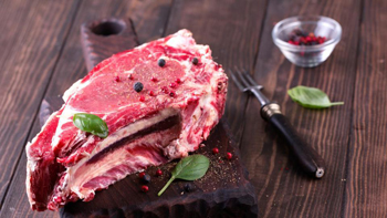
Washington, Jan 10: Men beware! Six servings a week of red meat, particularly the unprocessed variety, is said to heightened risk of developing common inflammatory bowel condition in men, warns a new study.

redmeatThe findings, published online in journal Gut, indicated that the highest level of red meat intake was associated with a 58% heightened risk of developing diverticulitis, with each daily serving associated with an 18% increased risk. However, risk peaked at six servings a week.

The association was strongest for unprocessed red meat and substituting one daily portion of this with fish or poultry was associated with a 20% lowered risk.

Diverticulitis occurs when the small pockets or bulges lining the intestine (diverticula) become inflamed.

The research team assessed the potential impact of total dietary red meat, poultry and fish intake on the risk of developing diverticulitis in nearly 46,500 men, aged 40 to 75.

They joined the study between 1986 and 2012 and after every four years they were asked to state how often, on average, they had eaten standard size portions of red meat, including processed meat; poultry; and fish, over the preceding year.

They were given nine options, ranging from ‘never’ or ‘less than once a month,’ to ‘six or more times a day.’

During the 26 year monitoring period, some 764 men developed diverticulitis.

But after taking account of these potentially influential factors, total red meat intake was associated with heightened diverticulitis risk.

But higher red meat consumption has been linked to the presence of inflammatory chemicals, such as C reactive protein and ferritin, as well as heart disease/stroke and diabetes, the researchers explained.

And the higher cooking temperatures involved for unprocessed meat, which was more strongly associated with diverticulitis, may influence bacterial composition or inflammatory activity, venture the researchers.

“Our findings may provide practical dietary guidance for patients at risk of diverticulitis, a common disease of huge economic and clinical burden,” the researchers concluded.

Comments

Add new comment

  • Coastaldigest.com reserves the right to delete or block any comments.
  • Coastaldigset.com is not responsible for its readers’ comments.
  • Comments that are abusive, incendiary or irrelevant are strictly prohibited.
  • Please use a genuine email ID and provide your name to avoid reject.
Agencies
June 29,2020

Washington DC, Jun 29: Young children with narrow retinal artery diameters were more likely to develop higher blood pressure, and children with higher blood pressure levels were more likely to develop retinal microvascular impairment during early childhood, according to a new study.

The first study to show this connection in children was published today in Hypertension, an American Heart Association journal.

High blood pressure, the main risk factor for the development of cardiovascular disease (CVD), can manifest as early as childhood, and the prevalence of high blood pressure among children continues to rise. In previous studies, analysis of blood vessels in the retina has shown promise as a predictor of CVD risk among adults. In the study titled, "Retinal Vessel Diameters and Blood Pressure Progression in Children," researchers sought to predict the development of high blood pressure in children over four years based on retinal blood vessel measurements.

"Hypertension continues as the main risk factor for the development of cardiovascular diseases and mortality," says Henner Hanssen, M.D., the study's lead author and a professor in the department of sport, exercise and health at the University of Basel in Switzerland. 

"Primary prevention strategies are needed to focus on screening retinal microvascular health and blood pressure in young children in order to identify those at increased risk of developing hypertension. The earlier we can provide treatment and implement lifestyle changes to reduce hypertension, the greater the benefit for these children."

Researchers screened 262 children ages six to eight from 26 schools in Basel, Switzerland, in 2014, for baseline blood pressure and retinal arterial measurements. Both measures were taken again in 2018. Blood pressure measurements at both baseline and follow-up were performed in a sitting position after a minimum of five minutes of rest and were categorized based on the American Academy of Pediatrics' blood pressure guidelines. These guidelines utilize the same measurements as the American Heart Association/American College of Cardiology 2017 Guideline for the Prevention, Detection, Evaluation, and Management of High Blood Pressure in Adults.

Results from the analysis indicate: children with narrower retinal vessel diameters at baseline developed higher systolic blood pressure at follow-up; retinal vessel diameters could explain 29 -31 per cent of the changes in systolic blood pressure progression between 2014 and 2018; children with higher blood pressure levels at baseline developed significantly narrower arteriolar diameters at follow-up, depending on weight and cardiorespiratory fitness; and initial blood pressure measures explained 66-69 per cent of the change in retinal arteriolar diameter from baseline to follow-up.

"Early childhood assessments of retinal microvascular health and blood pressure monitoring can improve cardiovascular risk classification. Timely primary prevention strategies for children at risk of developing hypertension could potentially counteract its growing burden among both children and adults," said Hanssen.

Researchers noted limitations of their study include that they could not confirm blood pressure measurements over a single 24-hour period, so they would not account for "white coat" hypertension, a condition where patients have high blood pressure readings when measured in a medical setting.

Developmental stage including puberty status of each child was not accounted for in the study, as well as genetic factors or birth weight - variables that could impact blood pressure development and microvascular health.

In addition, reference values for appropriate retinal vessel diameters in children do not currently exist, so future studies are needed to determine age-related normal values during childhood.

Comments

Add new comment

  • Coastaldigest.com reserves the right to delete or block any comments.
  • Coastaldigset.com is not responsible for its readers’ comments.
  • Comments that are abusive, incendiary or irrelevant are strictly prohibited.
  • Please use a genuine email ID and provide your name to avoid reject.
Agencies
May 14,2020

COVID-19 mostly kills through an overreaction of the immune system, whose function is precisely to fight infections, say scientists who have decoded the mechanisms, symptoms, and diagnosis of the disease caused by the SARS-Cov-2 coronavirus.

In a study published in the journal Frontiers in Public Health, the researchers explained step-by-step how the virus infects the airways, multiplies inside cells, and in severe cases causes the immune defences to overshoot with a "cytokine storm".

This storm is an over-activation of white blood cells, which release too-great amounts of cytokines -- inflammation-stimulating molecules --into the blood, they said.

"Similar to what happens after infection with SARS and MERS, data show that patients with severe COVID-19 may have a cytokine storm syndrome," said study author Daishun Liu, Professor at Zunyi Medical University in China.

"The rapidly increased cytokines attract an excess of immune cells such as lymphocytes and neutrophils, resulting in an infiltration of these cells into lung tissue and thus cause lung injury," Liu said.

The researchers explained that the cytokine storm ultimately causes high fever, excessive leakiness of blood vessels, and blood clotting inside the body.

It also causes extremely low blood pressure, lack of oxygen and excess acidity of the blood, and build-up of fluids in the lungs, they said.

The researchers noted that white blood cells are misdirected to attack and inflame even healthy tissue, leading to failure of the lungs, heart, liver, intestines, kidneys, and genitals.

This multiple organ dysfunction syndrome (MODS) may worsen and shutdown the lungs, a condition called acute respiratory distress syndrome, (ARDS), they said.

This, the researchers explained, happens due to the formation of a so-called hyaline membrane -- composed of debris of proteins and dead cells -- lining the lungs, which makes absorption of oxygen difficult.

Most deaths due to COVID-19 are therefore due to respiratory failure, they said.

The researchers explained that in the absence of a specific antiviral cure for COVID-19, the goal of treatment must be to the fight the symptoms, and lowering the mortality rate through intensive maintenance of organ function.

For example, an artificial liver blood purification system or renal replacement therapy can be used to filter the blood through mechanical means, they said.

The team noted that especially important are methods to supplement or replace lung function, for example with non-invasive mechanical ventilation through a mask, ventilation through a tube into the windpipe, the administration of heated and humidified oxygen via a tube in the nose, or a heart-lung bypass.

The researchers stressed the importance of preventing secondary infections.

They noted that SARS-Cov-2 also invades the intestines, where it causes inflammation and leakiness of the gut lining, allowing the opportunistic entry of other disease-causing microorganisms.

The researchers advocate that this should be prevented with nutritional support, for example with probiotics -- beneficial bacteria that protect against the establishment of harmful ones -- and nutrients and amino acids to improve the immune defences and function of the intestine.

"Because treatment for now relies on aggressive treatment of symptoms, preventative protection against secondary infections, such as bacteria and fungi, is particularly important to support organ function, especially in the heart, kidneys, and liver, to try and avoid further deterioration of their condition," Liu added.

Comments

Add new comment

  • Coastaldigest.com reserves the right to delete or block any comments.
  • Coastaldigset.com is not responsible for its readers’ comments.
  • Comments that are abusive, incendiary or irrelevant are strictly prohibited.
  • Please use a genuine email ID and provide your name to avoid reject.
News Network
February 21,2020

Washington, Feb 21: The fat around arteries may play an important role in keeping the blood vessels healthy, according to a study in rats that may affect how researchers test for treatments related to plaque buildup, as seen in conditions leading to heart attack.

The study, published in the journal Scientific Reports, noted that the fat, known as perivascular adipose tissue, or PVAT, helps arteries let go of muscular tension while under constant strain.

According to the researchers, including Stephanie W. Watts from the Michigan State University in the US, this feature is similar to how the bladder expands to accommodate more liquid, while at the same time keeping it from spilling out.

"In our study, PVAT reduced the tension that blood vessels experience when stretched," Watts said.

"And that's a good thing, because the vessel then expends less energy. It's not under as much stress," she added.

According to Watts and her team, PVAT has largely been ignored by researchers believing its main job was to store lipids and do little more.

Until now, she said, scientists only divided blood vessels into three parts, the innermost layer called the tunica intima, the middle layer called the tunica media, and the outermost layer called the tunica adventitia.

Watts believes PVAT is the fourth layer, which others have called tunica adiposa.

Tunica, she said, meant a membranous sheath enveloping or lining an organ, and adiposa is a synonym for fat.

"For years, we ignored this layer -- in the lab it was thrown out. In the clinic it wasn't imaged. But now we're discovering it may be integral to our blood vessels," Watts said.

"Our finding redefines what the functional blood vessels are, and is part of what can be dysfunctional in diseases that afflict us, including hypertension. We need to pay attention to this layer of a blood vessel because it does far more than we originally thought," she added.

Earlier studies, Watts said, had shown that PVAT plays a role in the functioning of blood vessels, finding that it secretes substances that can cause blood vessels to relax as well as substances that can cause it to contract.

In the current study, the researchers decided to test whether PVAT provides a structural benefit to arteries by assisting the function of stress relaxation.

They tested the thoracic aorta in rats, and found those with intact PVAT had more stress relaxation than those without.

The study revealed that the pieces of artery with surrounding fat had measurably relaxed more than those without.

Watts and her colleagues then tested other arteries, and were able to duplicate the same response.

"It's not something you see only in this particular vessel or this particular species or this particular strain. But that maybe it's a general phenomenon," she said.

Comments

Add new comment

  • Coastaldigest.com reserves the right to delete or block any comments.
  • Coastaldigset.com is not responsible for its readers’ comments.
  • Comments that are abusive, incendiary or irrelevant are strictly prohibited.
  • Please use a genuine email ID and provide your name to avoid reject.