Fat in poop linked with early presence of colorectal cancer

October 27, 2016

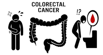
colorectalOct 27: Paving the way for a fast, noninvasive method that could lead to the early diagnosis of colorectal cancer, researchers have identified a suite of molecules in faeces that signifies the presence of precancerous polyps. ‘The exciting part is being able to see differences in the stool,’ said one of the researchers Herbert Hill, Professor at Washington State University. ‘This could lead to a noninvasive, more comprehensive early-warning detection method for colorectal cancer, but a lot of research needs to be done before it can be actually realised,’ Hill noted. Colorectal cancer is the second most common cancer worldwide. Nearly 1.4 million new cases were diagnosed in 2012, according to the World Cancer Research Fund International. Though early detection is key to successful treatment, most screening tests are limited in diagnostic capability or ease of application. Colonoscopy, for example, is a known lifesaver but is costly and unappealing to many people who might otherwise undergo testing. More people would be willing to provide a stool sample than undergo a biopsy through a colonoscope, Michael Williams from Washington State University, noted.

In addition, colonoscopes can only extend a limited distance into the large intestine, potentially missing some polyps. ‘With our new test, it could be possible to diagnose cancer occurring throughout the entire colon,’ he said. The researchers discovered the molecular fingerprint for colon cancer using a technology called ion mobility-mass spectrometry. They first identified metabolic products from normal colon tissue in both humans and mice. The scientists then compared this normal profile to that found in cancerous colon tissues from humans and research mice with polyps in their colons that mimic those in humans. In both cases, the scientists found that colon cancer caused significant changes in fat metabolism, especially for lipids and fatty acids. These abnormalities created a molecular fingerprint that was similar in humans and mice, said Hill. Next, the researchers examined droppings from transgenic and control mice to see if the molecular fingerprint could be found in feces as well. Here is how vitamin D helps to fight colorectal cancer.

Comments

Add new comment

  • Coastaldigest.com reserves the right to delete or block any comments.
  • Coastaldigset.com is not responsible for its readers’ comments.
  • Comments that are abusive, incendiary or irrelevant are strictly prohibited.
  • Please use a genuine email ID and provide your name to avoid reject.
Agencies
April 4,2020

Washington D.C., Apr 4: While consuming a high-diet salt can result in high blood pressure, a recent study has revealed a link between salt-rich diet and weaker immune system.

The study was conducted under the leadership of the University Hospital Bonn, and the results were published in the journal Science Translational Medicine.

The research was conducted on mice that were fed a high-salt diet. Later, they were found to suffer from much more severe bacterial infections.

Human volunteers who consumed an additional six grams of salt per day also showed pronounced immune deficiencies.

The World Health Organization (WHO) has recommended a maximum amount of five grams of salt a day.

It corresponds approximately to one level teaspoon. In reality, however, many Germans exceed this limit considerably. 

Figures from the Robert Koch Institute suggest that on average men consume ten, and women more than eight grams a day.

This means that we reach for the salt shaker much more than is good for us. After all, sodium chloride, which is its chemical name, raises blood pressure and thereby increases the risk of heart attack or stroke.

"We have now been able to prove for the first time that excessive salt intake also significantly weakens an important arm of the immune system," said Prof. Dr. Christian Kurts from the Institute of Experimental Immunology at the University of Bonn.

This finding is unexpected, as some studies point in the opposite direction. For example, infections with certain skin parasites in laboratory animals heal significantly faster if these consume a high-salt diet.

The study also sheds light on the fact that the skin serves as a salt reservoir.

"Our results show that this generalization is not accurate," emphasized Katarzyna Jobin, lead author of the study.

The body keeps the salt concentration in the blood and in the various organs largely constant. Otherwise important biological processes would be impaired. The only major exception is the skin which functions as a salt reservoir of the body. This is why the additional intake of sodium chloride works so well for some skin diseases.

However, other parts of the body are not exposed to the additional salt consumed with food. Instead, it is filtered out by the kidneys and excreted in the urine.

"We examined volunteers who consumed six grams of salt in addition to their daily intake," said Prof. Kurts. This is roughly the amount contained in two fast-food meals, i.e. two burgers and two portions of French fries.

After one week, from the results, it showed that the immune cells coped much worse with bacteria after the test subjects had started to eat a high-salt diet.

In human volunteers, excessive salt intake also resulted in increased glucocorticoid levels.

Comments

Add new comment

  • Coastaldigest.com reserves the right to delete or block any comments.
  • Coastaldigset.com is not responsible for its readers’ comments.
  • Comments that are abusive, incendiary or irrelevant are strictly prohibited.
  • Please use a genuine email ID and provide your name to avoid reject.
News Network
February 21,2020

Washington, Feb 21: The fat around arteries may play an important role in keeping the blood vessels healthy, according to a study in rats that may affect how researchers test for treatments related to plaque buildup, as seen in conditions leading to heart attack.

The study, published in the journal Scientific Reports, noted that the fat, known as perivascular adipose tissue, or PVAT, helps arteries let go of muscular tension while under constant strain.

According to the researchers, including Stephanie W. Watts from the Michigan State University in the US, this feature is similar to how the bladder expands to accommodate more liquid, while at the same time keeping it from spilling out.

"In our study, PVAT reduced the tension that blood vessels experience when stretched," Watts said.

"And that's a good thing, because the vessel then expends less energy. It's not under as much stress," she added.

According to Watts and her team, PVAT has largely been ignored by researchers believing its main job was to store lipids and do little more.

Until now, she said, scientists only divided blood vessels into three parts, the innermost layer called the tunica intima, the middle layer called the tunica media, and the outermost layer called the tunica adventitia.

Watts believes PVAT is the fourth layer, which others have called tunica adiposa.

Tunica, she said, meant a membranous sheath enveloping or lining an organ, and adiposa is a synonym for fat.

"For years, we ignored this layer -- in the lab it was thrown out. In the clinic it wasn't imaged. But now we're discovering it may be integral to our blood vessels," Watts said.

"Our finding redefines what the functional blood vessels are, and is part of what can be dysfunctional in diseases that afflict us, including hypertension. We need to pay attention to this layer of a blood vessel because it does far more than we originally thought," she added.

Earlier studies, Watts said, had shown that PVAT plays a role in the functioning of blood vessels, finding that it secretes substances that can cause blood vessels to relax as well as substances that can cause it to contract.

In the current study, the researchers decided to test whether PVAT provides a structural benefit to arteries by assisting the function of stress relaxation.

They tested the thoracic aorta in rats, and found those with intact PVAT had more stress relaxation than those without.

The study revealed that the pieces of artery with surrounding fat had measurably relaxed more than those without.

Watts and her colleagues then tested other arteries, and were able to duplicate the same response.

"It's not something you see only in this particular vessel or this particular species or this particular strain. But that maybe it's a general phenomenon," she said.

Comments

Add new comment

  • Coastaldigest.com reserves the right to delete or block any comments.
  • Coastaldigset.com is not responsible for its readers’ comments.
  • Comments that are abusive, incendiary or irrelevant are strictly prohibited.
  • Please use a genuine email ID and provide your name to avoid reject.
Agencies
July 24,2020

Colorado, Jul 24: A new study has found that physical stress in one's job may be associated with faster brain ageing and poorer memory.

Aga Burzynska, an assistant professor in the Department of Human Development and Family Studies, and her research team connected occupational survey responses with brain-imaging data from 99 cognitively normal older adults, age 60 to 79. They found that those who reported high levels of physical stress in their most recent job had smaller volumes in the hippocampus and performed poorer on memory tasks. The hippocampus is the part of the brain that is critical for memory and is affected in both normal ageing and in dementia.

Their findings were published this summer in Frontiers in Human Neuroscience under the research topic 'Work and Brain Health Across the Lifespan.'

"We know that stress can accelerate physical ageing and is the risk factor for many chronic illnesses," Burzynska said. "But this is the first evidence that occupational stress can accelerate brain and cognitive ageing."

She added that it is important to understand how occupational exposures affect the ageing of our brains.

"An average American worker spends more than eight hours at work per weekday, and most people remain in the workforce for over 40 years," Burzynska said. "By pure volume, occupational exposures outweigh the time we spend on leisure social, cognitive and physical activities, which protect our ageing minds and brains."

Physical demands at work

Burzynska explained that the association between "physical stress" and brain/memory were driven by physical demands at work. These included excessive reaching, or lifting boxes onto shelves, not necessarily aerobic activity. This is important because earlier work by Burzynska and her colleagues showed that leisure aerobic exercise is beneficial for brain health and cognition, from children to very old adults. Therefore, the researchers controlled for the effects of leisure physical activity and exercise.

As expected, leisure physical activity was associated with greater hippocampal volume, but the negative association with physical demands at work persisted.

"This finding suggests that physical demands at work may have parallel yet opposing associations with brain health," Burzynska explained. "Most interventions for postponing cognitive decline focus on leisure, not on your job. It's kind of unknown territory, but maybe future research can help us make some tweaks to our work environment for long-term cognitive health."

She added that the results could have important implications for society.

"Caring for people with cognitive impairment is so costly, on economic, emotional and societal levels," Burzynska said. "If we can support brain health earlier, in middle-aged workers, it could have an enormous impact."

The researchers considered and corrected for several other factors that could be related to work environment, memory and hippocampus, such as age, gender, brain size, educational level, job title, years in the occupation and general psychological stress.

One piece of the puzzle

"The research on this topic is so fragmented," Burzynska said. "One previous study linked mid-life managerial experience with greater hippocampus volume in older age. Another showed that taxi drivers had larger hippocampi than a city's bus drivers, presumably due to the need to navigate. In our study, job complexity and psychological stress at work were not related to hippocampal volume and cognition. Clearly, our study is just one piece of the puzzle, and further research is needed."

The magnetic resonance imaging (MRI) data used for the study was collected at the University of Illinois Urbana-Champaign between 2011 and 2014.

CSU researchers now can collect MRI data with the new 3T scanner at the University's Translational Medicine Institute.

With this new capability, Burzynska, along with Michael Thomas and Lorann Stallones of CSU's Department of Psychology, is launching a new project, "Impact of Occupational Exposures and Hazards on Brain and Cognitive Health Among Aging Agricultural Workers," which will involve collecting MRI brain scans and identifying risk and protective factors that could help the agricultural community age successfully. The project recently obtained funding as an Emerging Issues Short-Term Project from the High Plains Intermountain Center for Agricultural Health and Safety.

The Department of Human Development and Family Studies is part of CSU's College of Health and Human Sciences.
Comments

Add new comment

  • Coastaldigest.com reserves the right to delete or block any comments.
  • Coastaldigset.com is not responsible for its readers’ comments.
  • Comments that are abusive, incendiary or irrelevant are strictly prohibited.
  • Please use a genuine email ID and provide your name to avoid reject.