'Superbug from India resistant to all available antibiotics'

January 16, 2017

Washington, Jan 16: A woman in the US died after being infected by a superbug during her visit to India, say doctors who found that the "nightmare" bacteria was resistant to all available antibiotics.

bacteriaThe infection was caused by carbapenem-resistant Enterobacteriaceae (CRE), a multidrug-resistant organism associated with high mortality.

While CRE are not new to the US, what was new in this case is that the infection was resistant or non-susceptible to all available antimicrobial drugs, researchers said.

The 70-year-old patient in the US was admitted to an acute care hospital last year after an extended trip to India.

She was given a primary diagnosis of systemic inflammatory response syndrome, likely resulting from an infected right hip seroma. The infection was serious; none of the 14 antibiotics physicians used to treat the woman worked.

After the CRE - identified as Klebsiella pneumoniae - was confirmed by lab testing, an isolate from a wound specimen was sent to the CDC for further susceptibility testing and to determine the mechanism of resistance.

That testing confirmed the presence of New Delhi metallo-beta-lactamase (NDM-1), an enzyme that directly breaks down carbapenems, a powerful class of antibiotics that are often used to treat multidrug-resistant infections.

The US Centres for Disease Control and Prevention's antimicrobial testing showed the isolate was resistant to 26 different antibiotics, including all aminoglycosides and polymixins - another class of last-resort antibiotics.

It was also intermittently resistant to tigecycline, an antibiotic developed specifically to overcome drug-resistant organisms. Essentially, there were no treatment options.

Meanwhile, the patient's condition was deteriorating quickly. She died of septic shock in early September last year, less than two months after admission. The woman had been in India for two years and had been hospitalised at least four times there for a right femur fracture and a subsequent hip fracture.

"We feel comfortable saying that she most likely obtained the bug in India," said Lei Chen, senior epidemiologist with the Washoe County Health District in the US.

CRE have been labelled as a "nightmare" bacteria not only because they are already resistant to most antibiotics, but also because they spread easily in hospital settings.

The enzymes that enable resistance are carried on mobile pieces of DNA known as plasmids that can spread to other types of bacteria. NDM-1 enzyme is particularly mobile, researchers said.

"It's what we call a promiscuous plasmid, that seems to move easily between species," said Alex Kallen, a CDC medical epidemiologist.

When CRE bacteria enter the bloodstream, they can be deadly. CRE bacteria kill up to half of patients who get bloodstream infections from them.

NDM-1 was originally identified in 2009 in a Swedish patient who had been hospitalised in India.

The research was published in CDC's Morbidity and Mortality Weekly Report (MMWR).

Comments

Add new comment

  • Coastaldigest.com reserves the right to delete or block any comments.
  • Coastaldigset.com is not responsible for its readers’ comments.
  • Comments that are abusive, incendiary or irrelevant are strictly prohibited.
  • Please use a genuine email ID and provide your name to avoid reject.
Dr G K Sudhakar Reddy
August 4,2020

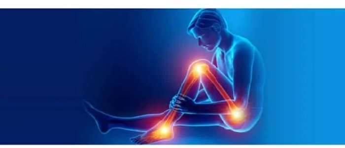
Being overweight or obese is now recognised as a serious cause of ill health and disability. There is a significant positive association between orthopaedic disorders and the level of obesity causing pain, deformity and difficulty in walking.

Excess body weight accumulation increases pressure on joints, particularly the hips, knees and ankles.

Here are a few type of  arthritis:

Osteoarthritis

It is a condition of damage/ wear and tear of the joint lining or cartilage. Obesity triggers this by loading excessive weight on the weight bearing joints like the knee and the hip. 

Knee Osteoarthritis

This is the most common arthritis especially in the Indian subcontinent.

While walking, an individual exerts 3 to 6 times pressure that of the body weight on the weight-bearing knee joint, which means in an obese with excess body weight, larger forces are exerted, which lead to higher risk of deterioration of cartilage.

In addition, there are excessive fat tissues that produce hormones and other factors that affect the joint cartilage metabolism and cause inflammation of the joints giving rise to joint pathology.  Leptin is one of the hormones causing knee osteoarthritis. 

Hip osteoarthritis

The force exerted across the hip is 3 times that of body weight. Hip osteoarthritis is caused by factors such as joint injury, increasing age and being overweight.    

Hand osteoarthritis

The observation that obese individual has a higher risk in having hand osteoarthritis has led to a hypothesis that the metabolic effect produced by fat tissue is the underlying factor. 

Osteoporosis

It is a progressive bone condition of decrease in bone mass and density (Bone Mineral Density or BMD) which can lead to an increased risk of fracture. Recent research suggests that obesity may accelerate bone loss. It is the amount of muscle mass which is seen in an active person, which accounts for bone strengthening effects and not due to the fat seen in a heavy person.

Low back pain

Low back pain from degenerative disc disease of the lumbar spine is one of the most disabling conditions in the community and overweight and obesity have the strongest association with seeking care for low back pain.

Managing Hip and Knee Osteoarthritis

Life style changes

If one is overweight, try to lose weight by doing more physical activity and eating a healthier diet. Regular exercise keeps you active and mobile and builds up muscle, thereby strengthening the joints and can improve symptoms. 

Pain Killers

Painkillers help with pain and stiffness for short term. They don’t affect the arthritis itself and won’t repair the damage to your joint. Creams and gels can be applied directly onto painful joints.

Nutritional Supplements

Glucosamine and chondroitin are nutritional supplements. Animal studies have found that glucosamine can both delay the breakdown of and repair damaged cartilage. However, there is insufficient evidence to support the use of glucosamine in humans and one can expect only a mild-to-moderate reduction in pain

Joint injections

If pain from osteoarthritis is severe joint steroid injections are injected into the joints that can reduces swelling and pain. The injections can start working within a day or so and may improve pain for several weeks or months. 

Hyaluronic acid injections, which help to lubricate your knee joint also give short term relief. In early stages. Stem cell treatment or cartilage regeneration procedures are being tried in young people with small defects, however it is still experimental and lacks long term evidence.

Surgery

May be recommended if you have severe pain or mobility problems.

Arthroscopy

If one has frequent painful locking/stiffening episodes especially in the knee joint, an operation to wash out loose fragments of bone and other tissue as joint can be performed by a minimally invasive key hole procedure called Arthroscopy.

Arthrodesis

If hip or knee replacement is not suitable, especially in young people who do heavy manual work, one can consider an operation known as an arthrodesis, which fuses your joint in a permanent position. This means that your joint will be stronger and much less painful, although you will no longer be able to move it.

Osteotomy

In young, active people in whom a knee joint replacement would fail due to excessive use one can consider an operation called an osteotomy. This involves adding or removing a small section of bone either above or below your knee joint.  This helps realign your knee so your weight is no longer focused on the damaged part of your knee. An osteotomy can relieve your symptoms of osteoarthritis, although you may still need knee replacement surgery eventually as you grow old

Joint replacement surgery

Joint replacement therapy is most commonly carried out to replace hip and knee joints. It involves replacing a damaged, worn or diseased joint with an artificial joint made of special plastics and metal.

For most people, a replacement hip or knee will last for at least 20 years, especially if it is cared for properly and not put under too much strain.

Dr G K Sudhakar Reddy is a Sr Consultant Orthopaedic Surgeon at Citizens Speciality Hospitals, Hyderabad

Comments

Add new comment

  • Coastaldigest.com reserves the right to delete or block any comments.
  • Coastaldigset.com is not responsible for its readers’ comments.
  • Comments that are abusive, incendiary or irrelevant are strictly prohibited.
  • Please use a genuine email ID and provide your name to avoid reject.
News Network
February 21,2020

Washington, Feb 21: The fat around arteries may play an important role in keeping the blood vessels healthy, according to a study in rats that may affect how researchers test for treatments related to plaque buildup, as seen in conditions leading to heart attack.

The study, published in the journal Scientific Reports, noted that the fat, known as perivascular adipose tissue, or PVAT, helps arteries let go of muscular tension while under constant strain.

According to the researchers, including Stephanie W. Watts from the Michigan State University in the US, this feature is similar to how the bladder expands to accommodate more liquid, while at the same time keeping it from spilling out.

"In our study, PVAT reduced the tension that blood vessels experience when stretched," Watts said.

"And that's a good thing, because the vessel then expends less energy. It's not under as much stress," she added.

According to Watts and her team, PVAT has largely been ignored by researchers believing its main job was to store lipids and do little more.

Until now, she said, scientists only divided blood vessels into three parts, the innermost layer called the tunica intima, the middle layer called the tunica media, and the outermost layer called the tunica adventitia.

Watts believes PVAT is the fourth layer, which others have called tunica adiposa.

Tunica, she said, meant a membranous sheath enveloping or lining an organ, and adiposa is a synonym for fat.

"For years, we ignored this layer -- in the lab it was thrown out. In the clinic it wasn't imaged. But now we're discovering it may be integral to our blood vessels," Watts said.

"Our finding redefines what the functional blood vessels are, and is part of what can be dysfunctional in diseases that afflict us, including hypertension. We need to pay attention to this layer of a blood vessel because it does far more than we originally thought," she added.

Earlier studies, Watts said, had shown that PVAT plays a role in the functioning of blood vessels, finding that it secretes substances that can cause blood vessels to relax as well as substances that can cause it to contract.

In the current study, the researchers decided to test whether PVAT provides a structural benefit to arteries by assisting the function of stress relaxation.

They tested the thoracic aorta in rats, and found those with intact PVAT had more stress relaxation than those without.

The study revealed that the pieces of artery with surrounding fat had measurably relaxed more than those without.

Watts and her colleagues then tested other arteries, and were able to duplicate the same response.

"It's not something you see only in this particular vessel or this particular species or this particular strain. But that maybe it's a general phenomenon," she said.

Comments

Add new comment

  • Coastaldigest.com reserves the right to delete or block any comments.
  • Coastaldigset.com is not responsible for its readers’ comments.
  • Comments that are abusive, incendiary or irrelevant are strictly prohibited.
  • Please use a genuine email ID and provide your name to avoid reject.
Agencies
July 3,2020

Apart from the many benefits of doing exercise, new research has now found that exercise can slow down or prevent the development of macular degeneration and may benefit other common causes of vision loss, such as glaucoma and diabetic retinopathy.

The new study from the University of Virginia School of Medicine found that exercise reduced the harmful overgrowth of blood vessels in the eyes of lab mice by up to 45 per cent. This tangle of blood vessels is a key contributor to macular degeneration and several other eye diseases.

The study represents the first experimental evidence showing that exercise can reduce the severity of macular degeneration, a leading cause of vision loss, the scientists report. Ten million Americans are estimated to have the condition.

"There has long been a question about whether maintaining a healthy lifestyle can delay or prevent the development of macular degeneration. The way that question has historically been answered has been by taking surveys of people, asking them what they are eating and how much exercise they are performing," said researcher Bradley Gelfand, PhD, of UVA's Center for Advanced Vision Science.

"That is basically the most sophisticated study that has been done. The problem with that is that people are notoriously bad self-reporters ... and that can lead to conclusions that may or not be true. This [study] offers hard evidence from the lab for the very first time," Gelfand added.

Enticingly, the research found that the bar for receiving the benefits from exercise was relatively low - more exercise didn't mean more benefit.

"Mice are kind of like people in that they will do a spectrum of exercise. As long as they had a wheel and ran on it, there was a benefit. The benefit that they obtained is saturated at low levels of exercise," Gelfand said.

An initial test comparing mice that voluntarily exercised versus those that did not found that exercise reduced the blood vessel overgrowth by 45%. A second test, to confirm the findings, found a reduction of 32 per cent.

The scientists aren't certain exactly how exercise is preventing the blood vessel overgrowth. There could be a variety of factors at play, they say, including increased blood flow to the eyes.

Gelfand, of UVA's Department of Ophthalmology and Department of Biomedical Engineering, noted that the onset of vision loss is often associated with a decrease in exercise.

"It is fairly well known that as people's eyes and vision deteriorate, their tendency to engage in physical activity also goes down. It can be a challenging thing to study with older people. ... How much of that is one causing the other?" he said.
The researchers already have submitted grant proposals in hopes of obtaining funding to pursue their findings further.

"The next step is to look at how and why this happens, and to see if we can develop a pill or method that will give you the benefits of exercise without having to exercise," Gelfand said.

He explained, "We're talking about a fairly elderly population [of people with macular degeneration], many of whom may not be capable of conducting the type of exercise regimen that may be required to see some kind of benefit." (He urged people to consult their doctors before beginning any aggressive exercise program.)
Gelfand, a self-described couch potato, disclosed a secret motivation for the research: "One reason I wanted to do this study was sort of selfish. I was hoping to find some reason not to exercise," he joked. "It turned out exercise really is good for you."

Comments

Add new comment

  • Coastaldigest.com reserves the right to delete or block any comments.
  • Coastaldigset.com is not responsible for its readers’ comments.
  • Comments that are abusive, incendiary or irrelevant are strictly prohibited.
  • Please use a genuine email ID and provide your name to avoid reject.